The central dogma of Biology is DNA –> RNA –> protein. But how do we get there? This is where the processes of transcription and translation come into play. Transcription is the process by where RNA is synthesized from a DNA template and translation is the process by where protein is made from the RNA (mRNA) template. Let’s discuss in detail, how each process works.
Transcription
In Eukaryotic cells, transcription occurs in the nucleus. DNA is double stranded, with one strand running 5’ –> 3’ and the other running parallel at 3’ –> 5’. Transcription or RNA synthesis, involves the DNA strand running 3’ –> 5’ or the DNA template. RNA polymerase, an enzyme needed for this process, binds to the promotor region of the DNA. As RNA polymerase moves down and opens up the DNA, it adds nucleotides based on the DNA code. The nucleotides that are added are base pairs to the DNA template. The RNA polymerase will move down until it encounters a stop codon, at which point the RNA will be released. The RNA that is released is actually pre-mRNA. It contains both introns and exons. In order to be properly read and turned into protein, the introns need to be spliced out by RNA splicing and the exons put together. Once this process happens, the mature mRNA, will exit the nucleus through the nuclear pores and enter into the cytoplasm.
Translation
Once the mRNA is in the cytoplasm, it can bind to a ribosome where the process of translation will take place. In order for translation to occur, there are three RNA’s needed. These are the ribosome or rRNA, the mRNA which binds to the ribosomes and just came from the nucleus, and tRNA. mRNA is read in triplets or codons, so every three nucleotides. tRNA has anticodons, so three nucleotides that will match with the codons on the mRNA. For each anticodon tRNA has, it carries a specific amino acid. So again, the amino acid that tRNA carries is based on its anticodon. This allows the proteins that each mRNA codes for to be made the same way every time. tRNA travels to the ribosome, binds its anticodon to mRNAs codon and deposits its amino acid, this will continue until the entire amino acid chain (polypeptide) is made and the tRNA reaches a stop codon. (This process is much more specific and I have explained it thoroughly in the video I linked below) At this point, the protein will be released. It can be folded, have posttranslational modifications made to it, be chaperoned somewhere. At this point the protein is made and what happens next just really depends on its function.
*NOTE: In prokaryotic cells (cells which have no nucleus), transcription and translation both occur in the cytoplasm, usually at the same time.
Here is a video that I made on this topic. It goes into much more detail so that you can really grasp this concept:
Here is a study guide / fun fact sheet I made on this topic:
Let me know what you think. Should I make more fact sheets? Do you like the video? Comment below and let me know your thoughts or if you have any questions.
COVID-19 (SARS-CoV-2), a novel coronavirus, which transferred to humans in late December 2019. Originally, referred to as Wuhan Coronavirus as the first cases of patients seem to be associated with the Huanan seafood market or surrounding area in Wuhan, China. This virus causes viral pneumonia with the most prominent symptoms being cough, fever and difficulty breathing1. The radiographs of most patients with the virus show invasive lesions in both lungs. However, the symptoms can vary from mild to severe with some patients being asymptomatic. The incubation period of the virus can last anywhere from 2-14 days, according to the CDC which is basing it off the MERS-CoV viruses1. The Wuhan Coronavirus was previously being referred to as 2019-nCoV, but is now being called COVID-19.
This virus is part of the family Coronaviridae. The Coronaviridae family of viruses are normally seen in birds, bats, camels, masked palm civets, mice, dogs and cats. Normally, these viruses are kept within the avian or mammalian species it infects. However, occasionally, a spillover event occurs in which the virus is transferable to humans. Of the coronaviruses that are pathogenic to humans, most are either mild or asymptomatic2. The exceptions to these are SARS, Severe acute respiratory syndrome, and MERS, Middle East respiratory syndrome. SARS was identified in November 2002, Guangdong, China3. SARS causes severe respiratory illness and during the 2002-2003 outbreak, infected over 8000 people and caused 774 deaths3. MERS was identified in Saudia Arabia in 2012. The symptoms of MERS cover a full spectrum of cases from severe disease, to mild, to asymptomatic4. Symptoms can include fever, runny nose, cough, with the common factor being viral pneumonia.
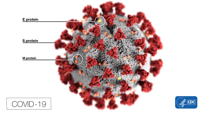
This leads us to our new novel finding of the Wuhan Coronavirus, COVID-19. COVID-19 has been identified to be in the same family, Coronaviridae, as SARS and MERS. We are still discovering new properties of this novel virus, but let’s take a look at what we do know. The Coronaviridae family of viruses are enveloped, single-stranded, positive-sense RNA viruses. They are subdivided into four groups: alpha, beta, gamma and delta, of which SARS, MERS and COVID-19 fall into the beta group. They contain very large genomes for an RNA virus, approximately 30 kilobases (kb). Coronaviridae’s virion structure is spherical with diameters about 125nm. The clublike projections that protrude from the surface give the viruses a solar corona appearance, leading to the name coronavirus. These viruses contain an envelope with a nucleocapsid inside. The nucleocapsids for these viruses are symmetrical which is uncharacteristic of positive-sense strand RNA viruses, this a common feature for negative-sense strand RNA viruses. There are four main structural proteins found in these viruses, encoded for on the 3’ end of their genome: spike (S), membrane (M), envelope (E), and nucleocapsid (N)5. Inside the nucleocapsid is where you would find the protected genome of the virus.
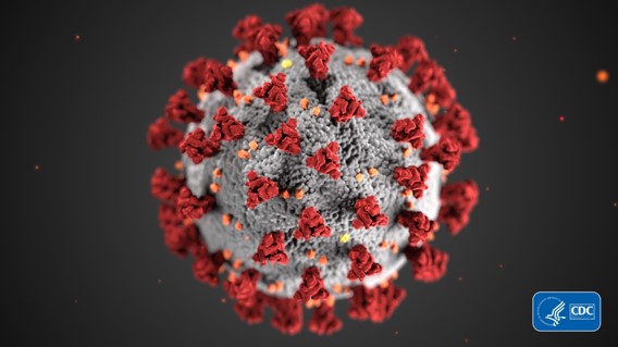
Recent images created by the CDC show the solar corona appearance of the virus when viewed with the electron microscope.
*images from CDC image libraryRecent images created by the CDC show the solar corona appearance of the virus when viewed with the electron microscope.
*images from CDC image library
For a coronavirus to enter into a cell, it requires the S protein to come into contact with its receptor5. The S protein/receptor binding is a key component in the ability of the coronavirus to infect a host, and it also determines what cell type(s) it can infect. Once bound to its receptor, the virus must gain access into the cell’s cytosol. This occurs through a series of cleavages of the S protein, which allows the virus and cellular membranes to fuse. Following this fusion, the viral genome is released into the cell’s cytoplasm
Once the viral genome has entered into the cytoplasm, the next step is for the replicase gene to be translated from the genome. Once this gene is translated, many cleavages occur that result in the formation of the replicase-transcriptase complex (RTC) which is responsible for RNA replication and transcription. After replication and transcription of the viral particles occurs, assembly and release can follow. The M protein is responsible for directing most of the protein-protein interaction in the virus construction. It has been suggested that the M protein together with the E protein are responsible for producing the coronavirus envelopes5,6. Once the virions have finished assembly, they are transported to the cell surface in vesicles and released by exocytosis. It has been suggested that the E protein is not only involved in viral assembly, but also in viral release7. (*All of these processes’ have been simplified and are much more complex involving various other steps. If you would like to read more about it please see paper #5 in the Bibliography).
What receptor is COVID-19 using to infect cells? Recently, it has been shown that COVID-19 is using ACE2 receptors to attach to and infect primary host lung cells8,9. Since COVID-19 seems to use ACE2 receptors to enter into the host cell, it is of interest to know which cells contain these receptors. Knowing where these receptors are expressed and distributed, would give some insight into the infection routes of the virus. One location where ACE2 receptors are highly expressed is type II alveolar cells, which are found in the lungs11-13. Other cells with ACE2 highly expressed are cells in the esophagus (upper and stratified), absorptive enterocytes in the ileum and colon13, cholangiocytes (bile duct), myocardial cells, kidney proximal tubule cells and bladder and urothelial cells11. In addition, Xu et al., just recently discovered that ACE2 is expressed in the oral cavity epithelial cells, with higher expression in the tongue than buccal and gingival tissues10.
The best way to prevent the spread of COVID-19 is to make sure your hands are washed, avoid touching your face and avoiding contact with others that may be sick. Those who are more at risk include immune comprised, older adults and those who may have underlying health conditions, such as diabetes, heart or lung disease.
2. Su S, Wong G, Shi W, et al. Epidemiology, genetic recombination, and pathogenesis of coronaviruses. Trends Microbiol 2016; 24: 490–502.
3. Peiris JS, Guan Y, Yuen KY. Severe acute respiratory syndrome. Nat Med 2004; 10 (suppl 12): S88–97.
4. Hui DS, Azhar EI, Kim Y-J, Memish ZA, Oh M-d, Zumla A. Middle East respiratory syndrome coronavirus: risk factors and determinants of primary, household, and nosocomial transmission. The Lancet Infectious Diseases. 2018;18(8):e217-e27.
5. Fehr AR, Perlman S. Coronaviruses: an overview of their replication and pathogenesis. Methods Mol Biol. 2015;1282:1–23. doi:10.1007/978-1-4939-2438-7_1
6. Bos EC, Luytjes W, van der Meulen HV, Koerten HK, Spaan WJM. The production of recombinant infectious DI-particles of a murine coronavirus in the absence of helper virus. Virology. 1996; 218:52–60.
7. Ye Y, Hogue BG. Role of the coronavirus E viroporin protein transmembrane domain in virus assembly. Journal of virology. 2007; 81(7):3597–3607.
8. Yushun Wan, Jian Shang, Rachel Graham, Ralph S. Baric, Fang Li. Receptor recognition by novel coronavirus from Wuhan: An analysis based on decade-long structural studies of SARS. Journal of Virology Jan 2020, JVI.00127-20; DOI: 10.1128/JVI.00127-20
10. Xu, H., Zhong, L., Deng, J. et al. High expression of ACE2 receptor of 2019-nCoV on the epithelial cells of oral mucosa. Int J Oral Sci12, 8 (2020). https://doi.org/10.1038/s41368-020-0074-x
11. Zou, X. et al. The single-cell RNA-seq data analysis on the receptor ACE2 expression reveals the potential risk of different human organs vulnerable to Wuhan 2019-nCoV infection. Front. Med. http://journal.hep.com.cn/fmd/EN/ 10.1007/s11684-020-0754-0 (2020).
12. Zhao, Y. et al. Single-cell RNA expression profiling of ACE2, the putative receptor of Wuhan 2019-nCov. Preprint at https://www.biorxiv.org/content/10.1101/ 2020.01.26.919985v1 (2020).
13. Zhang, H. et al. The digestive system is a potential route of 2019-nCov infection: a bioinformatics analysis based on single-cell transcriptomes. Preprint at https:// http://www.biorxiv.org/content/10.1101/2020.01.30.927806v1 (2020).
Basic explanation of antigens and antibodies and how they interact. More in-depth discussions on this topic will happen at a later time.
What is an antigen (Ag)?
Substance (molecule) that the body recognizes as foreign
Substance capable of provoking an immune response
Substance that stimulates the immune system to make antibodies
Example: Virus, bacteria
Recap: An antigen is a foreign substance that the body does not recognize as one of its own, as such, the body is capable of engaging in an immune response against it. This idea is more complex and will be further discussed in a later blog/video.
What is an antibody (Ab)?
Specialized protein that can help immune cells destroy antigens
Also referred to as Immunoglobulins (proteins found in the blood)
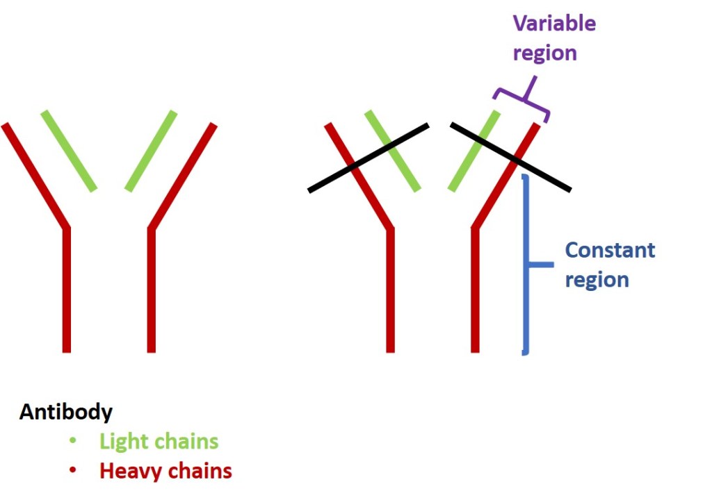
Composed of 2 Heavy and 2 Light chains (refer to figure below)
Composed of a Constant and Variable regions (refer to figure below)
Constant region – remains the same for all antibodies. Antibodies that are attached to cell would have this region embedded in the cell membrane
Variable region – as the name implies, this region is variable. This region is also where an antibody would bind an antigen. Since this region varies, it allows for antibodies to be able to bind various antigens. This topic is more complex and will be discussed in more detail in a later blog/video.
Recap: An antibody is a specialized protein, made by the body, capable of binding to various antigens.
How do antibodies and antigens interact?
An antibody binds an antigen in its variable site or antigen binding site. This binding blocks the antigen from doing harm to the body and flags it for removal. For example, if the antigen was a virus, the antibody would bind to the virus and now the virus can no longer infect cells. This is one of the many defense systems the body uses against foreign substances.
*Note, there are different types of antibody classes and different functions of antibodies, these will be discussed in later blog/videos. This is only a basic explanation of antibodies and antigens to help those who may be struggling with the concept.
There are several words that you have probably seen that are used to describe homeostasis:
Equilibrium
Stability
Balance
Steady state
Basically, living organisms need to be able to maintain balance within their internal environment, especially compared to the outside environment. Imagine going outside on an incredibly hot or cold day and your internal temperature were to change to maintain balance with the external environment. This would not be healthy and would upset everything in the body. As such, there are mechanisms put into place so that this does not happen.
These mechanisms include:
Negative feedback – this mechanism works to reduce or reverse a change in the internal environment
Positive feedback – this mechanism amplifies a change or keeps it going in the same direction.
Feedback systems are how biological processes can self-regulate or maintain stability (homeostasis). Most of the control mechanisms of homeostasis are based on negative feedback, which is the most common form of regulation in living systems. However, though less common, there are also biological processes that are regulated by positive feedback systems.
Negative Feedback
This common mechanism works to reverse a change. When the normal “set points” are disrupted, these mechanisms can kick in and bring the change back to the normal set point. For example, when you go outside and it is freezing, your body keeps on mechanisms to keep your internal temperature constant. These mechanisms reverse the change (your body being cold outside) and work to keep internal temperature constant. There are many examples of negative feedback which you will cover throughout your course. For now, I will include a couple of examples here for you to understand it better.
Figure showing two examples of Negative feedback. These mechanisms reverse the changes of either the temperature rising or decreases and bring the body temperature back to it’s set point.
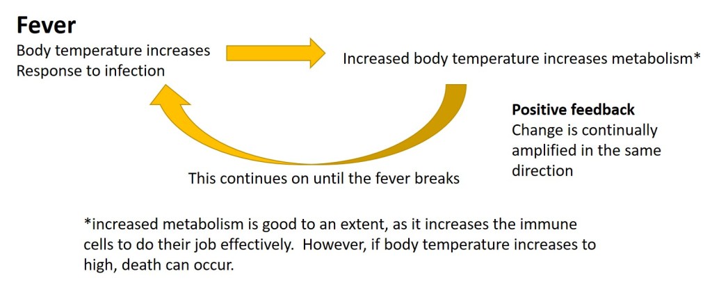
Positive Feedback
These mechanisms are not as common, but do exist. Rather than working to reverse a change in order to maintain homeostasis, these mechanisms amplify a change and keep the change going in the same direction. I will include a couple of examples below for you to understand it better.
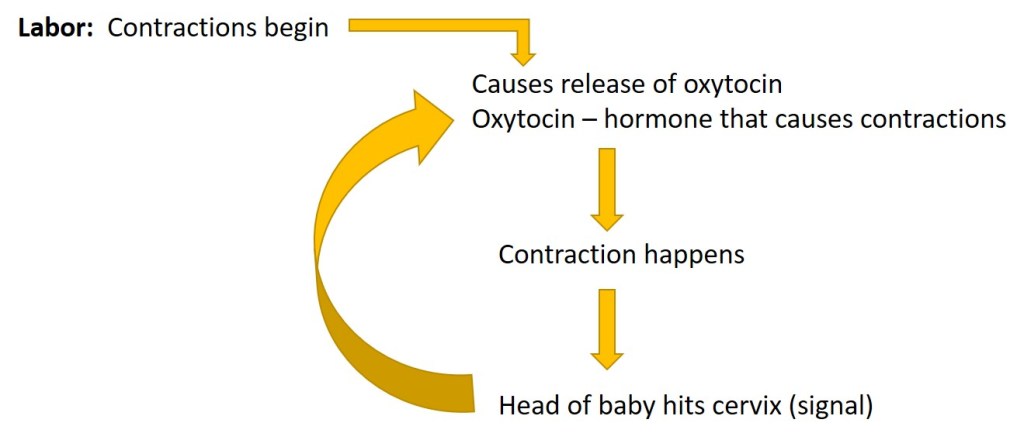
This type of feedback, amplifies the change and keeps it going in the same direction. Once signal is gone (baby is born). Positive feedback mechanism signals will start to subside.Another example of positive feedback.
If you would rather hear the explanations of these feedback systems or have a little more in-depth discussion of this topic, please check out my YouTube video at:
If you like my video, please make sure to subscribe to my channel so you are always kept up to date on new video additions, and like and share the video. Thank you so much.


You must be logged in to post a comment.